
Write something

4d •
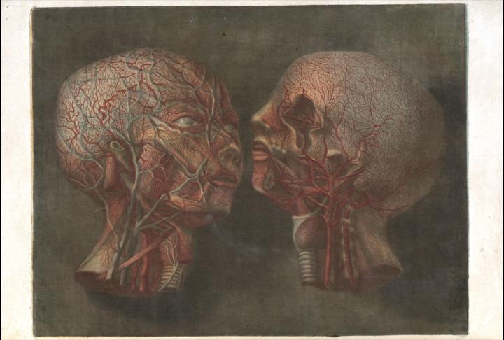
Anatomie de la tête (1748)
Anatomie de la tête (1748) is one of those works that forces you to think about what we are actually doing when we “look” at the human body. In 18th-century Paris, anatomy was establishing itself as a modern science, but learning it still relied on a practical problem: access to the body and to live demonstrations. Enter Jacques Fabien Gautier d’Agoty, an artist and printing technician who understood that a good image could be an instrument of knowledge. His gamble was massive: to publish a series of large, life-size plates—eight in total—focusing on the human head. We are not talking about "little drawings for a manual," but images made to impose themselves, ensuring the student (or the artist) would want to stay and observe. Gautier d’Agoty collaborated with surgeon Joseph-Guichard Duverney, who was linked to anatomical teaching in Paris and was responsible for preparing the specimens and dissections; Gautier d’Agoty then translated that physical reality into a printed image. The foundation, despite what it might seem, is not an idealized imagination, but a visual record of a real anatomical preparation. The work shows, for example, cranial dissections where the skull is opened to reveal internal structures, with special attention paid to the visual impact of what is exposed. The artistic quality of these pieces cannot be understood without the technique: color mezzotint. Unlike black-and-white prints that were hand-colored afterward, here color is part of the printing process itself. This allows for richer transitions, deeper shadows, and a sense of volume that could not be achieved before. This is where the figure of Jacob Christoph Le Blon appears, associated with the development of color printing, which Gautier d’Agoty utilized and pushed into the anatomical realm. That is why these plates function more as visual objects than mere "illustrations": they are designed to seduce the eye. It is fascinating that this atlas did not only compete with other atlases; it competed with the "experience of dissection" itself. In his dedication to Louis XV, Gautier d’Agoty defends the value of his "printed paintings" against the shock that direct dissection could cause. It is almost a pedagogical proposal: if the real body can be too violent or inaccessible, a well-crafted image can serve as a mediator.
2
0
6d •
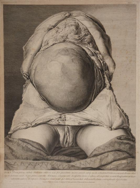
The Anatomy of the Human Gravid Uterus Exhibited in Figures (1774) / The Anatomy of the Human Gravid Uterus Exhibited in Figures. 1774
The Anatomy of the Human Gravid Uterus Exhibited in Figures (whose original Latin title is Anatomia uteri humani gravidi tabulis illustrata) is one of the crowning achievements in the history of medical illustration and obstetrics. It was published in 1774 by the Scottish physician and anatomist William Hunter (brother of the equally famous surgeon John Hunter), and it is celebrated both for its monumental contribution to the understanding of pregnancy and for its striking, hyper-realistic aesthetic. The creation of this atlas took more than 24 years of systematic research due to the enormous difficulty of acquiring the corpses of women who had died during advanced stages of gestation. William Hunter began the project in 1750, when his anatomy school in London obtained, for the first time, the body of a woman who had died at nine months pregnant. Unlike previous books, where the uterus was depicted from the outside in or based on imagination, Hunter decided to open the corpses and rigorously document the womb from the inside out to show the different stages of fetal development (from 5 weeks to 9 months). The most outstanding element of the book is its 34 breathtaking copperplate engravings. They were printed in a spectacular folio format (known as an elephant folio), which allowed the illustrations to be life-size (a 1:1 scale) representations of the anatomical specimens. The vast majority of the original drawings were made by the Dutch artist Jan van Rymsdyk, whom Hunter forced to draw directly over the dissection table, forbidding him from illustrating from memory or embellishing the scene. As a result, Rymsdyk's style is hyper-realistic and raw: the images show not only the anatomy of the fetus, placenta, and uterus, but also the "ugly" and real details of the environment, such as the irregular cuts of the skin, the folds of fat, pins, and even the wooden blocks or ropes that supported the corpse. Precisely because of that lack of polish, the book possesses a rare and overwhelming artistic power. Each plate carries a sense of strangeness and of an unrepeatable opportunity. It forces you to look at the body as evidence, not as an icon, and that honesty has a dark component, because it reminds you of the material price of that knowledge.
2
0
8d •
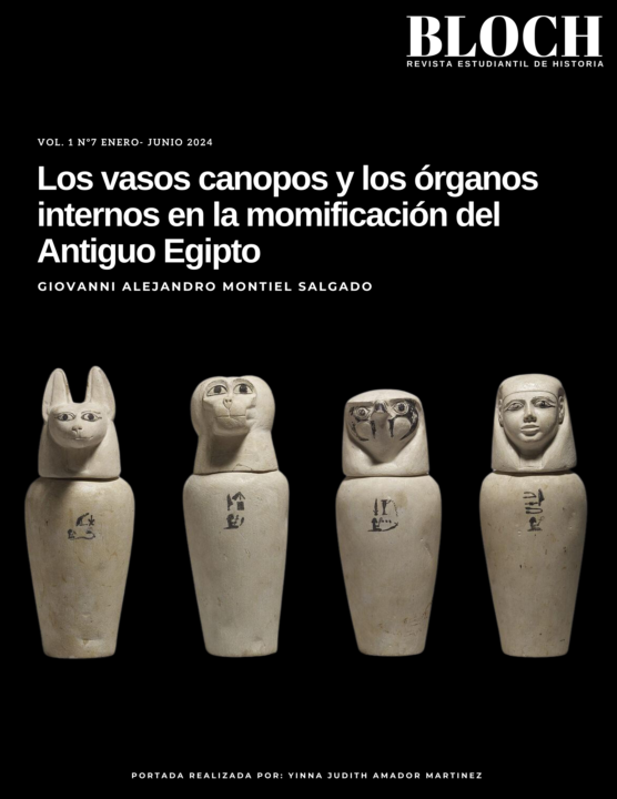
Why Did the Egyptians Throw the Brain in the Trash? / ¿Por qué los egipcios tiraban el cerebro a la basura?
We have been studying the human body for thousands of years, and for most of that time, we understood it in a very different way than we do now. This is no minor detail: the way a civilization conceives its own body is not just a scientific issue; it is the foundation upon which it builds its culture, its morality, and its worldview. Every boundary it draws between the accessory and the functional, between the physical and the spiritual, ultimately shapes its institutions, its language, and the experience its individuals have of themselves. The body, in short, is not just the object of study: it is the map through which all reality is interpreted. For centuries in ancient Egypt, embalmers eviscerated bodies with enormous precision. They knew how to separate the liver from the gallbladder, recognized major fluid canalization systems, and developed specialized instruments. However, when preparing the bodies, they did something that, from our modern perspective, seems bizarre: they extracted the brain through the nostrils with a metal hook and discarded it along with the rest of the useless tissues and fluids. Conversely, they left the heart carefully intact in its place: it was the only organ that was untouched and not stored in a canopic jar. Why did they do this? To answer, it is helpful to first understand who the embalmers really were and what place they occupied in Egyptian society, because their role was very different from that of a physician. Physicians (swnw) enjoyed enormous social prestige, usually came from wealthy families, and trained in the prestigious "Houses of Life," attached to the temples. They were scholars who combined empirical clinical observation with pharmacological treatments and rituals. Embalmers, on the other hand, worked in mummification workshops—sometimes called "Houses of Death"—and belonged to a priestly class specializing in funerary rites. Their manual labor with corpses, the handling of corrosive liquids, and the smells associated with putrefaction placed them in a social category considered impure or repellent to the general population.
2
0
1-3 of 3

skool.com/representar-la-anatomia
A multidisciplinary community dedicated to exploring anatomy, art, technology, and aesthetics. illuminating the dark architecture of the flesh.
Powered by