Activity
Mon
Wed
Fri
Sun
May
Jun
Jul
Aug
Sep
Oct
Nov
Dec
Jan
Feb
Mar
Apr
What is this?
Less
More
Owned by Juan
A multidisciplinary community dedicated to exploring anatomy, art, technology, and aesthetics. illuminating the dark architecture of the flesh.
Memberships
DESPIERTA TU EROS
5 members • Free
Skoolers
195k members • Free
13 contributions to Depicting Anatomy (EN/ES)
28d •
📌 Temporary schedule update: Recorded classes and upcoming live sessions
I apologize for the sudden change, but due to unforeseen circumstances, this week's classes will not be held live. Live sessions will resume starting April 7th. Until then, I will continue uploading the recorded classes according to our schedule so you can watch them at your convenience. Please feel free to leave any questions you might have in the comments section of each video or in the community forum, and I will get back to you as soon as possible. Thank you very much for your understanding. ------ 🗓️ Aviso importante: Actualización sobre las clases en directo Siento comunicaros que, por motivos imprevistos, las sesiones de esta semana no podrán realizarse en directo. Las clases en vivo se reanudarán a partir del 7 de abril. Hasta entonces, seguiré subiendo las sesiones grabadas a la plataforma respetando el calendario previsto, para que podáis avanzar a vuestro propio ritmo. Por favor, dejad cualquier duda que os surja en los comentarios de cada vídeo o en el foro de la comunidad, y os responderé lo antes posible. Agradezco mucho vuestra comprensión.
3
0
Mar 18 •
🚨 A New Era Begins! (And a lifetime gift for those already here) 🚨
Hi everyone! I know the community has been a bit quiet these past few weeks, and I apologize for that. The truth is, I've been working hard behind the scenes, preparing something massive. I wanted to take this space to the next level so it becomes the best place on the internet to master anatomical representation... and everything is finally ready! To sustain this pace, content quality, and dedication, the community is transitioning to a paid model. BUT WAIT: If you are reading this inside the group, it means you trusted this project from the beginning. And that deserves a reward. That's why, all current members are automatically upgraded to the Founder Tier. This means you will have access to all Premium tier benefits completely free, forever (as long as you don't leave the community). It's my way of saying thank you for being here. Starting next week, we are going all out. Get ready because we are going to sculpt, model, and learn like never before. Here is a breakdown of what each tier includes from now on: 🛡️ Founder Tier (Standard) The exclusive tier for the pioneers. As a current member, you get all the Premium tier benefits automatically and forever, without paying anything. (You only lose it if you decide to leave the community). ⚡ Premium Tier: The engine of your progress How will the community work from now on? Your Premium access gives you a steady workflow to take your portfolio to the next level: 🔴 2 Weekly Live Sessions (45 min each) We will have rotating themes covering all angles of representation: - Sectional Anatomical Studies: In-depth analysis of specific regions (bones, muscles, tension) to understand their real mechanics and volume. - Technical Workflows: Modeling in ZBrush, texturing in Substance Painter, and rendering in Blender. - Historical Anatomy and Analysis: We study how masters like Vesalius or Da Vinci resolved the body to apply it to our work today. - Live Reviews: Professional and constructive critiques of the work uploaded by the community.
1 like • Mar 18
@Luise Köhne Thank you. I really want to make this community an extraordinary place. It’s the exact hub I would have loved to be a part of when I started my journey in anatomical representation. I have a lot of great plans and ideas ahead for us. Now it’s time to put in the work and make this space a reality.
Mar 18 •
💸 New Affiliate System: Earn Money by Recommending the Community
I’m excited to announce something new: from now on, you’ll also be able to earn money by recommending the community to people who can truly benefit from it. The idea is simple: if you know artists, illustrators, sculptors, modelers, or students who want to improve their anatomical representation skills, you’ll be able to invite them using your personal affiliate link and earn a commission for every person who joins through you. This is not about selling for the sake of selling. It’s about recommending something you know, use, and genuinely believe can help other artists grow. How does it work? - You will receive your own personal affiliate link. - You share that link with people you believe would benefit from the community. - If someone joins through your link, you earn a commission. - The more qualified people you invite, the more you can earn. Why am I doing this? Because this community has always grown better through real recommendations than through cold advertising. People who join because someone inside recommended it usually come in with more clarity, commitment, and motivation to do the work. And honestly, I think it’s only fair that if you help grow this space, you should benefit from it too. How much can you earn? - Commission for every new member who joins through your link: 40% Who should you invite? This works best if you invite people who are genuinely a good fit for the community: - Artists who want to improve anatomy for illustration or 3D. - People who need structure, feedback, and consistency. - Anyone interested in ZBrush, Blender, anatomical representation, and the history of anatomy. - Artists who want to take their portfolio to a more professional level. Important I’m not looking for spam or aggressive promotion. I would much rather have one honest recommendation than ten mass messages with no context. If you share the community, do it with intention: explain why you think this space could help the other person and what they can actually find inside. That way, everyone wins.
2
0
Mar 13 •
Something important is about to change...
Depicting Anatomy is about to enter a new phase. More ambitious, more refined, and much more aligned with the core vision that gave meaning to this community from the very beginning. It has taken me a little time to shape it, and I deeply appreciate your patience. I am not going to share the exact changes just yet, but I do want to give you one certainty: current members will hold a very special place, and your trust will be rewarded. All the details will be revealed next week. Thank you for being here before everything changes. I hope what is to come excites you as much as it does me. If you have any suggestions or something you would love to see, please don't hesitate to leave them in the comments below. 👇 --- Algo importante está a punto de cambiar... Representar la anatomía está a punto de entrar en una nueva fase. Más ambiciosa, más cuidada y mucho más alineada con la visión de fondo que dio sentido a esta comunidad desde el principio. Me ha llevado un poco de tiempo darle forma y os agradezco enormemente vuestra paciencia. Aún no voy a compartir los cambios, pero sí quiero dejar una certeza: los miembros actuales ocuparéis un lugar muy especial, y vuestra confianza tendrá recompensa. La próxima semana se revelarán todos los detalles. Gracias por estar aquí antes de que todo cambie. Espero que lo que está por venir os emocione tanto como a mí. Si tenéis alguna sugerencia o algo que os encantaría ver, no dudéis en dejarla en los comentarios. 👇
0 likes • Mar 13
@Earth Ki You are the foundation of this community. I´m here for you.
Mar 9 •
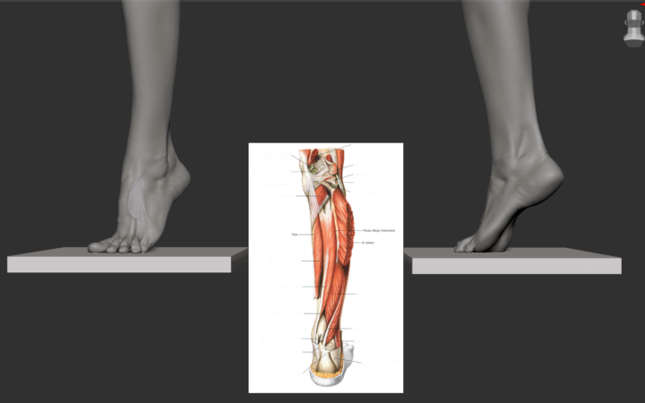
🎨 New project underway! Anatomical model of the foot and lower leg in ZBrush.
Hey community! Today I start sculpting this new anatomical model, featuring all the complexity of a dissected piece. I'll record the entire creation process. The end result will be a complete tutorial so you can see my ZBrush workflow step by step. I'll post progress updates, timelapse clips, and specific tips. What interests you most: muscle modeling, proportions, references, the final finish...? Comment below and let's learn together! 💪🦴 #ZBrush #Anatomy #DigitalSculpting #Tutorial --- 🎨 ¡Nuevo proyecto en marcha! Modelo anatómico del pie y parte de la pierna en ZBrush. ¡Hola comunidad! Hoy empiezo la escultura de este nuevo modelo anatómico, con toda la complejidad de una pieza diseccionada. Voy a grabar el proceso de creación. El resultado será un tutorial completo para que veáis mi flujo de trabajo en ZBrush paso a paso. Subiré avances, clips del timelapse y tips específicos. ¿Qué parte os interesa más: el modelado de los musculos, las proporciones, el acabado final? ¡Comentad abajo y vamos aprendiendo juntos! 💪🦴 #ZBrush #Anatomia #EsculturaDigital #Tutorial
4
0
1-10 of 13

@juan-caso-8679
Fundador de Anatomy One. +15 años en edición médica creando modelos 3D para arte y cirugía. Ayudo a comunicar la anatomía con rigor y visión artística
Active 1d ago
Joined Feb 24, 2026
Madrid
Powered by