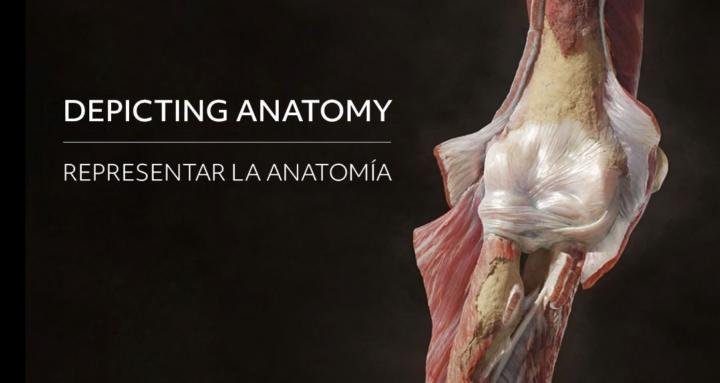
(Or Why Your Sculpts Look Like Action Figures)
Take a stroll through ArtStation or Sketchfab searching for "human anatomy," and you'll spot the pattern: hundreds of technically flawless models that, nevertheless, look completely artificial, superheroic, hyper-inflated. They look like 90s Marvel action figures.
The mistake is almost always the same: by trying to reconstruct internal anatomy—and marking it exaggeratedly to show off everything you know—you end up treating every muscle as if it were an independent balloon.
Most modelers learn anatomy by studying origins and insertions, and then they sculpt each muscle belly separately (the biceps, the brachioradialis, the quadriceps...). They stack them in layers, carving deep grooves between them and adding the volume of each one to whatever lies beneath.
The result might be "correct" in theory. You've placed the humerus, and over it the brachialis muscle, the coracobrachialis, and the deltoid covering the top... And they are all worked independently with their own volume and shape. We’ve all been there, but in practice, this shatters realism.
The Two Main Culprits: Fascia and Gravity
The underlying problem is that we ignore two key elements: fascia and gravity.
Fascia is the connective tissue that wraps and compresses the musculature. Imagine taking two tenderloins of meat, placing a stick between them, and wrapping them tightly with cling film. That is fascia. The muscles are compacted inside. In a living, relaxed body, muscles don't burst outward: they flatten against each other, they overlap, and gravity also pulls their mass downward.
When you sculpt a biceps, you are not just sculpting the biceps. You are sculpting the biceps, covered by the brachial fascia, covered by subcutaneous fat, and covered by skin. Those are four layers compressing, softening, and redistributing the volume.
The secret to anatomical realism is not adding more volume. It’s understanding compression. A relaxed muscle hangs. A muscle in tension flattens against the bone and the fascia containing it.
The Real Thicknesses:
In the table, you can see some examples of the real thicknesses measured by ultrasound and MRI in healthy adults at rest (the perpendicular distance between the superficial and deep aponeurosis). In many cases, the muscles are no thicker than a slice of ham. (Note: in elite athletes, these values can double).
The muscles that visually appear the most imposing—the pectoralis, the deltoid, the trapezius—are actually wide but flat. The perception of power comes from their spread, not from a spherical volume. The truly thick and dense masses lie deep: the vastus lateralis, the gastrocnemius, the glutes.
3 Tips to Improve Your Anatomy Work
- Appearance ≠ Volume.Before adding mass, ask yourself: does this muscle generate width or thickness? A well-sculpted pectoral is a shape that extends laterally and merges with the deltoid and the sternum, not a sphere protruding from the chest.
2. Respect the Fascia.The grooves between muscles exist, but they are soft. In a relaxed body, connective tissue and subcutaneous fat fill in and smooth out those transitions. Mark the separation just enough for it to read, and then smooth it out.
3. Think in Layers, Not in Pieces.The human body is not a model kit. Sculpt keeping in mind that the skin is the final result of everything underneath, not a wrapper you add at the end.
The Best References: MRIs, Cadavers, and Plastinations
No anatomy book for artists will give you the same information as a cadaveric cross-section. Seeing the actual cross-sectional area of a biceps or a quadriceps—exactly as it appears on an MRI—permanently recalibrates how you perceive muscle volume.
Plastinated figures, where body fluids are replaced by polymers, are especially valuable because they maintain the real thickness of the muscle in dynamic poses, showing the mechanical tension of the fibers without the shrinkage caused by formaldehyde.
As I said at the beginning, we have all made these mistakes during our learning process.
Do you want to share examples of your own, or ones you’ve found online, of super-inflated anatomies?
Negative examples are just as interesting as positive ones, if not more so.
I'll be reading you in the comments!
---
Deja de inflar los músculos (o por qué tus esculturas parecen figuras de acción)
Date una vuelta por ArtStation o Sketchfab buscando "human anatomy" y verás el patrón: cientos de modelos técnicamente impecables que, sin embargo, se ven completamente artificiales, superheroicos, hiperinflados. Parecen figuras de acción de Marvel de los 90.
El error casi siempre es el mismo: al intentar reconstruir la anatomía interna —y marcarla exageradamente para demostrar todo lo que sabes—, acabas tratando cada músculo como si fuera un globo independiente.
La mayoría de los modeladores aprenden anatomía estudiando orígenes e inserciones, y luego esculpen cada vientre muscular por separado (el bíceps, el braquiorradial, los cuádriceps...). Los van apilando en capas, marcando surcos profundos entre ellos y sumando el volumen de cada uno al que está debajo.
El resultado sería "correcto" en teoría. Has colocado el húmero, y sobre él el músculo braquial, el coracobraquial y el deltoides cubriendo por encima… Y todos trabajados de forma independiente con su volumen y su forma. Todos hemos pasado por ahí, pero en la práctica, esto rompe el realismo.
Los dos grandes culpables: la fascia y la gravedad
El problema de fondo es que ignoramos dos elementos clave: la fascia y la gravedad.
La fascia es el tejido conectivo que envuelve y comprime la musculatura. Imagina que coges dos solomillos de carne, colocas un palo entre ellos y los envuelves fuertemente con film transparente. Eso es la fascia. Los músculos quedan compactados dentro. En un cuerpo vivo y relajado, los músculos no estallan hacia afuera: se aplastan entre sí, se solapan, y además la gravedad tira de su masa hacia abajo.
Cuando esculpes un bíceps, no estás esculpiendo solo el bíceps. Estás esculpiendo el bíceps, cubierto por la fascia braquial, cubierto por la grasa subcutánea, y cubierto por la piel. Son cuatro capas que comprimen, suavizan y redistribuyen el volumen.
El secreto del realismo anatómico no es añadir más volumen. Es entender la compresión. Un músculo relajado cuelga. Un músculo en tensión se aplasta contra el hueso y la fascia que lo contiene.
Los grosores reales:
En la tabla, podéis ver algunos ejemplos de los grosores reales medidos por ecografía y resonancia magnética en adultos sanos en reposo (la distancia perpendicular entre la aponeurosis superficial y la profunda). En muchos casos, los músculos no son más gruesos que una loncha de jamón. (Nota: en atletas de élite, estos valores pueden duplicarse).
Los músculos que visualmente parecen más imponentes —el pectoral, el deltoides, el trapecio— son en realidad anchos pero planos. La percepción de potencia viene de su extensión, no de un volumen esférico. Las masas realmente gruesas y densas están en profundidad: el vasto lateral, el gastrocnemio, los glúteos.
3 consejos para mejorar tus trabajos de anatomía
1. Apariencia ≠ volumen.
Antes de añadir masa, pregúntate: ¿este músculo genera anchura o grosor? Un pectoral bien esculpido es una forma que se extiende lateralmente y se fusiona con el deltoides y el esternón, no una esfera que sobresale del tórax.
2. Respeta la fascia.
Los surcos entre músculos existen, pero son suaves. En un cuerpo relajado, el tejido conectivo y la grasa subcutánea rellenan y suavizan esas transiciones. Marca la separación apenas lo suficiente para que se lea, y luego suaviza.
3. Piensa en capas, no en piezas.
El cuerpo humano no es un kit de montaje. Esculpe pensando en la piel como el resultado final de todo lo que hay debajo, no como un envoltorio que añades al final.
Las mejores referencias: resonancias, cadáveres y plastinaciones
Ningún libro de anatomía para artistas te dará la misma información que un corte transversal cadavérico. Ver el área de sección real de un bíceps o un cuádriceps —exactamente como aparece en una resonancia magnética— recalibra de forma permanente cómo percibes el volumen muscular.
Las figuras plastinadas, donde los fluidos del cuerpo son reemplazados por polímeros, son especialmente valiosas porque mantienen el grosor real del músculo en poses dinámicas, mostrando la tensión mecánica de las fibras sin la retracción del formaldehído.
Como decía al principio, todos hemos cometido estos errores durante nuestro aprendizaje.
¿Quieres compartir ejemplos tuyos, o que hayas encontrado en internet, de anatomías súper infladas? Los ejemplos negativos son tan interesantes o más que los positivos.
¡Os leo en los comentarios!