

Apr 18 •
What I Wish I Knew Before Medical School
I went to medical school to become an orthopedic surgeon. I was ranked to match. And then I walked away from medicine entirely. In this video, I'm sharing everything I wish someone had told me before I started — the burnout, the opportunity cost, the reality of being locked into one specialty for decades, and what I'd do differently if I could go back. If you're a premed wondering whether medical school is right for you, or a med student having doubts about your path, this is the video I wish existed when I was in your shoes. 🔥 Want personalized 1-on-1 support from physician tutors? Apply for a strategy call with our DO-founded tutoring team: 👇 https://calendly.com/d/cyf5-dgv-6jc/tutoring-consultation-application-call
Mar 28 •
Not Every DO Student Needs a COMLEX Tutor… Do You?
Many tutoring companies suggest every DO student needs a tutor, but I'm here to offer a different perspective. Having personally tutored hundreds of DO students and scored in the 99th percentile on COMLEX, I want to be transparent about who truly benefits from a tutor and who doesn't. Most medical students don't need a private tutor if their scores are consistently above 450 and trending upwards, and often they just need to adjust their study plan. This insight into osteopathic medical education and med school tips aims to clarify common misconceptions about COMLEX tutoring and how to approach exam preparation effectively. 🔥 Want personalized 1-on-1 support from physician tutors? Apply for a strategy call with our DO-founded tutoring team: 👇 https://calendly.com/d/cyf5-dgv-6jc/tutoring-consultation-application-call
1
0
Mar 14 •
Why Your DO School is Wrong About USMLE
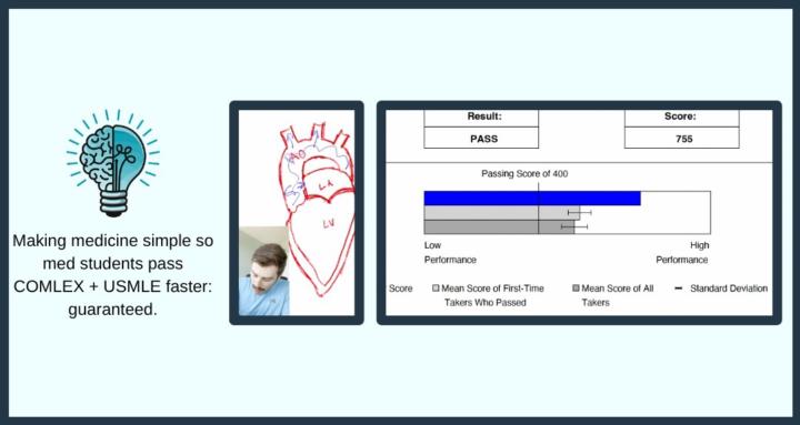
Most DO students don't actually need to take both COMLEX and USMLE. In this video, I explain exactly why. As a former medical school professor and current board prep educator, I break down when USMLE preparation makes sense and when it's an unnecessary burden for an osteopathic medical student. You'll learn how to decide if taking USMLE step 1 and 2 for a DO is truly worth it for your career path. 🔥 Want personalized 1-on-1 support from physician tutors? Apply for a strategy call with our DO-founded tutoring team: 👉 https://calendly.com/d/cyf5-dgv-6jc/tutoring-consultation-application-call
0
0
Mar 1 •
She Failed COMLEX Level 1...Then This Happened On Her Next Attempt
Shannon failed COMLEX Level 1 on her first attempt. Not because she wasn’t smart. Not because she didn’t work hard. Her biggest problem? Resource overload. Too many videos. Too many question banks. Too many flashcards. No clear system. After working with us for two months, everything changed. We simplified her study plan, fixed her question strategy, and focused on comprehension instead of drowning in content. Then… the unexpected happened. On test day, she locked her ID in her car and wasn’t allowed to sit for the exam. Another delay. Another setback. But she didn’t spiral. She stayed the course — and passed COMLEX Level 1 on her second attempt. In this interview, Shannon explains: • Why resource overload caused her first failure • What actually changed in her studying • How she stayed calm after the ID incident • What she would do differently from day one If you’re overwhelmed by COMLEX prep or worried about failing, this conversation is for you. Have questions for Shannon? Contact her here: sbkirkla2020@gmail.com 🔥 Want personalized 1-on-1 support from physician tutors? Apply for a strategy call with our DO-founded tutoring team: 👉 https://calendly.com/nrusso-premeducated/tutoring-consult Study less. Understand more. Pass your boards.
2
0
Feb 21 •
140 DO Med Students Asked Me These 5 Questions About Boards
An osteopathic medical school flew me out to lecture 140 DO medical students on COMLEX prep and over three days, I heard the same five questions again and again. If you’re preparing for COMLEX Level 1, chances are you’re asking them too. In this video, I break down: • When and how often to take COMSAEs • What to do if Sketchy doesn’t work for micro and pharm • Whether your dedicated period is long enough • Should you take USMLE and COMLEX — and which first? • How to study when your school forces you to attend class during “dedicated” We also talk about: • The 450 COMSAE benchmark and what it actually means • Why spaced repetition (Anki) is non-negotiable • How to structure questions vs content review • And the biggest misconception DO students have about boards I’m Dr. Lucas — former medical school professor, DO, and full-time COMLEX tutor. This is exactly what I told 140 osteopathic medical students before they started dedicated. If you’re staring down Level 1, this will save you time, money, and unnecessary panic. 🔥 Want personalized 1-on-1 tutoring built for DO and MD retakers? Book a strategy call with our team of physicians: 👉 https://calendly.com/nrusso-premeducated/30min Study less. Score higher. Touch grass.
2
0
1-15 of 15
skool.com/premeducated
Live COMLEX tutoring & study community built by DO physicians for DO students. Get answers, coaching, and accountability to raise your score quickly.
Leaderboard (30-day)
1

+3
2

+3
3

+3
4

+3
5
+3
Powered by