
Write something
Jun 8 •
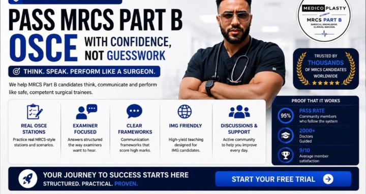
🩺 Medicoplasty MRCS Part B Accelerator
🎯 Promise Pass MRCS Part B with a structured, examiner-focused programme designed to eliminate overwhelm and maximise your chance of passing at the next sitting. 📚 What You Get ✅ Complete MRCS Part B curriculum ✅ Weekly live teaching sessions ✅ Weekly mock OSCE stations ✅ High-yield question bank ✅ Complete notes library ✅ Private accountability community ✅ Recorded sessions ✅ Personal study roadmap 🎁 Bonuses - Anatomy Image Bank - Lifetime access to recordings 🛡️ Pass Commitment Follow the programme, attend sessions, complete the work, and if you don't pass, you can join the next cohort free. 💰 Investment Normal Price £600 Founding Cohort Offer £400 Save £200 🔥 Why This Is Different Most MRCS courses give you information. Medicoplasty gives you a complete passing system: - What to study - How to study - When to study - Weekly accountability - Mock practice - Expert feedback - A community of candidates preparing with you 🚀 Outcome Walk into the MRCS Part B exam knowing exactly what examiners expect and having practised the stations repeatedly before exam day. Train Smarter. Pass Faster. Operate With Confidence. 🩺🚀 Limited Spaces Available for the End of June Cohort. Comment here or Send What's app text to 0447453315868
2
0

Jan 30 •
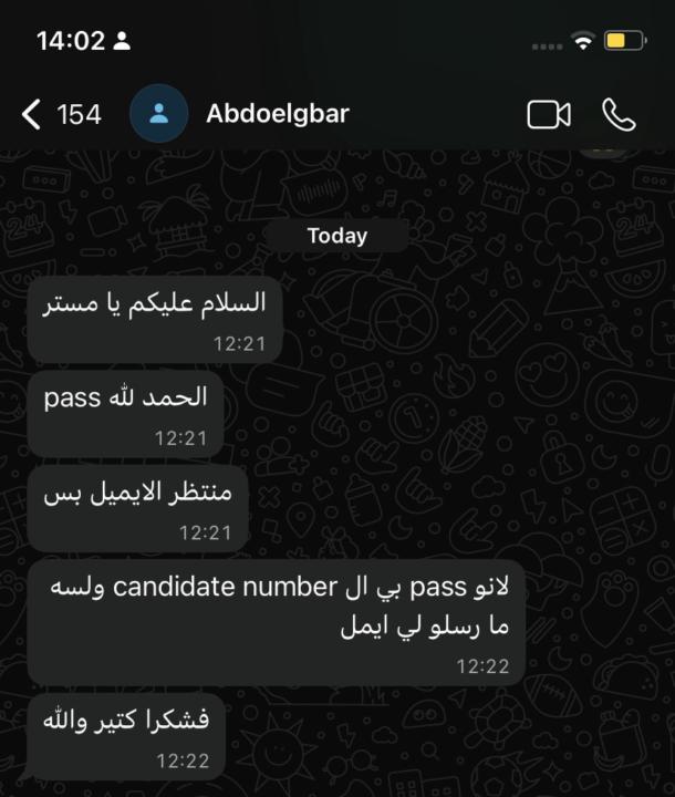
🎉 MRCS Part B Success! 🎉
I am pleased to congratulate Alaa Aljamri, Abdoalgbar, and Abubaker on passing the MRCS Part B examination today. Many congratulations to you all, and I wish you continued success in your careers and in life. To all our other candidates, I wish you every success in passing this examination.
1-30 of 44
powered by
Suggested communities
Powered by