
Activity
Mon
Wed
Fri
Sun
Jun
Jul
Aug
Sep
Oct
Nov
Dec
Jan
Feb
Mar
Apr
May
What is this?
Less
More
Memberships
MSK Radiology School
Public • 780 • Free
Pre-RadPodSquad
Private • 53 • Free
THP Jump Training
Private • 47.2k • Free
112 contributions to MSK Radiology School

10d ago
in
Phantom Coalition
Bilateral radiographs w/ weight-bearing and right ankle MRI performed https://www.cmrad.com/cases/1735748974 Patient was subsequently taken to the OR for a calcaneonavicular coalition. The issue is that I"m having trouble seeing it on the pre-operative images :S
1
18
New comment 2d ago
1 like • 6d
@Marlena Jbara Love the insight re: repairing plantar plate tears without addressing the cause
0 likes • 2d
@Im Beg Agreed, thanks for sharing!

0 likes • 15d
I concur Disclaimer: Only looked at the stills
15d ago
in
Intraosseous varix
Patient scanned for concern for hamstring pathology (can scroll through and try to identify where/which muscle is injured for bonus points) Incidentally noted lesion in the femur, no prior radiograph. Reported as an intraosseous varix. I think I agree but wanted to hear others' thoughts as well https://www.cmrad.com/cases/1241031294
1
1
New comment 15d ago

16d ago
in
Periostitis ?
40ish sports man 80 km a week complain of exquisit pain at inf 1/3 of right tibia . There is hypersignal of the tibialis anterior muscle . Is this a sign of perisotitis or a compartment syndrom ?
2
5
New comment 15d ago
2 likes • 16d
Based on the still images and clinical history, agree with traction periostitis from tibialis anterior This RadSource article has a nice discussion of differential. They talk about how differentials, including chronic exertional compartment syndrome (CECS) and bony stress injuries, can all coexist. One technique they describe for CECS is to perform imaging before and after exercise and to look for increased muscle size and edema after exercise As for other etiologies, the author adds muscle strain, DVT, muscle hernia, etc. DVT is an interesting consideration given the ancillary findings in your case: "However, unlike compartment syndrome, deep venous thrombosis causes venous occlusion and commonly results in subcutaneous edema and skin thickening, which are all findings that can be displayed with MR imaging" https://radsource.us/compartment_syndrome_leg/
18d ago
in
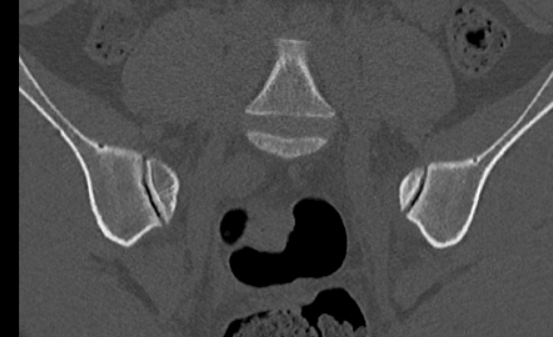
Low back pain
26 year Old Lady , no prior pregnancy . 3 months of all time ache . HLA B27 neg . Antélysthesis L5 S1 with bilatéral pars brake . Last image was done in 2021( abdominal CT for abdominal pain).
3
8
New comment 16d ago
1 like • 16d
Very difficult case, looking forward to the follow-up MRI. Thanks Professor Hermann for the explanation
1-10 of 112